Home
/ Tendon Diagram Leg - Diagram Showing The Tendons And Ligaments Of The Ankle And Foot Download Scientific Diagram / Included are more than a dozen illustrations like the vastus lateralis, adductor brevis, rectus femoris, semi.
Tendon Diagram Leg - Diagram Showing The Tendons And Ligaments Of The Ankle And Foot Download Scientific Diagram / Included are more than a dozen illustrations like the vastus lateralis, adductor brevis, rectus femoris, semi.
Tendon Diagram Leg - Diagram Showing The Tendons And Ligaments Of The Ankle And Foot Download Scientific Diagram / Included are more than a dozen illustrations like the vastus lateralis, adductor brevis, rectus femoris, semi.. (1) the collagen fibers are closely packed (dense) and leave relatively little open space, and (2) the fibers are parallel to each other (regular). This important tendon in the back of the calf and ankle stores the elastic energy needed for running. Both are made of collagen. The early and late management. Both of these types of structure may.
Included are more than a dozen illustrations like the vastus lateralis, adductor brevis, rectus femoris, semi. One of the most important tendons in terms of mobility of the leg is the achilles tendon. The carpal tunnel is a tube of nerves and tendons that passes through the wrist. This site contains information about tendon diagram of leg. If so, these resistance band leg stretches are going to benefit you greatly.
Plantaris Muscle Wikipedia from upload.wikimedia.org Anatomy of leg muscles and tendons anatomy diagram leg. This site contains information about tendon diagram of leg. Tendon diagram of calf and knee. If so, these resistance band leg stretches are going to benefit you greatly. A ligament is often found in the joints of the body and are connective fibrous tissues from bone to bone. Hip, thigh, leg & tendon muscle diagrams. Both of these types of structure may. This system works to provide both stability and mobility while we walk or run.
A tendon or sinew is a tough band of fibrous connective tissue that connects muscle to bone and is capable of withstanding tension.
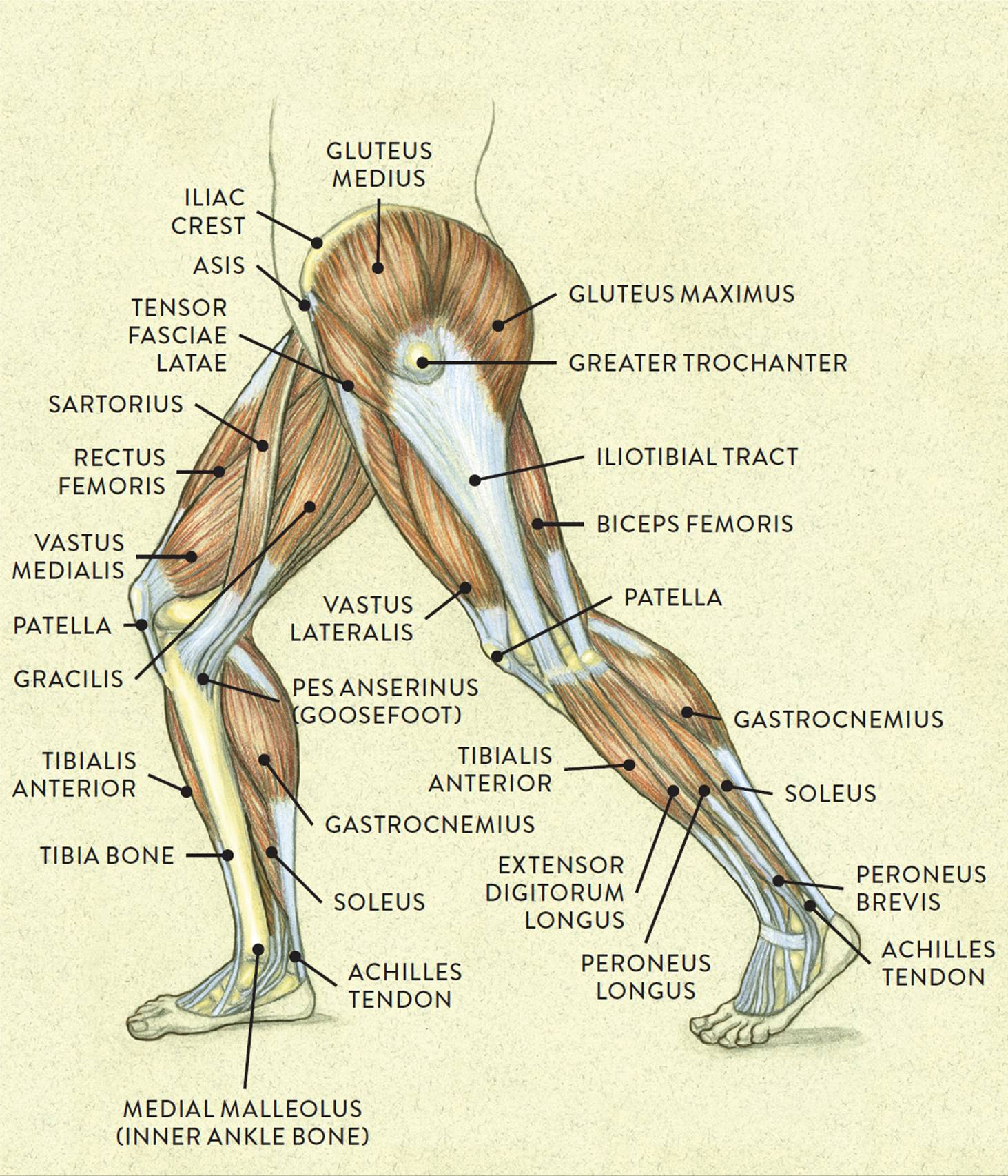
This diagram with labels depicts and explains the details of leg tendons anatomy. Hip, thigh, leg & tendon muscle diagrams. The carpal tunnel is a tube of nerves and tendons that passes through the wrist. The early and late management. A tendon or sinew is a tough band of fibrous connective tissue that connects muscle to bone and is capable of withstanding tension. Are your legs sore or tight? J bone joint surg br 69:416420 fig. (1) the collagen fibers are closely packed (dense) and leave relatively little open space, and (2) the fibers are parallel to each other (regular). Small sacs of fluid called bursae cushion the achilles tendon at the heel. Both tendons and ligaments are dense regular connective tissue, because of its two properties: Hip, thigh, leg & tendon muscle diagrams. Posted on january 21, 2015 by admin. Leg muscle and tendon diagram google search muscle.
Posted on january 21, 2015 by admin. Tendons are similar to ligaments; Tendon vs ligament medlineplus medical encyclopedia image. Tendon diagram muscle tendon diagram 9 out of 10 based on 40 ratings. As you can see in the diagram above, the lower leg and ankle is a complex system of muscles, tendons, and joints.
Muscles Of The Leg And Foot Classic Human Anatomy In Motion The Artist S Guide To The Dynamics Of Figure Drawing from doctorlib.info Posted on january 21, 2015 by admin. One of the most important tendons in terms of mobility of the leg is the achilles tendon. These sensors synapse on interneurons in the spinal cord that inhibit further activity of the motor neurons innervating the muscle. A tendon or sinew is a tough band of fibrous connective tissue that connects muscle to bone and is capable of withstanding tension. The horizontal lines and the percentages used to measure the distances of sural nerve and the small saphenous vein are marked on the cadaver. Each of these muscles is a discrete organ constructed of skeletal muscle tissue. Leg muscle and tendon diagram google search muscle, foot and ankle anatomical chart, diagram of list of wiring diagrams, foot anatomy bones ligaments muscles tendons arches, ligaments of the foot tendons in the foot wedding love. Superficial and deep anterior muscles of upper body.
There are two main muscle groups around the knee:
Knee tendon joint ligament anatomy foot medical muscle bone cartilage fibula illustration kneecap lateral leg movement structure tibia anatomical anterior athlete body cap care connect crucuate diagram education femur fitness graphic health health care human iliotibial isolated medial medicine. The carpal tunnel is a tube of nerves and tendons that passes through the wrist. Tendon diagram muscle tendon diagram 9 out of 10 based on 40 ratings. #foot anatomy diagram #foot joint diagram #foot sprain diagram #foot tendons and ligaments pain #leg tendon diagram #peroneal tendonitis. Human anatomy and physiology diagrams: Should the alignment of the foot and leg be out the foot muscles are forced to work harder to compensate which only works to a tendon back of knee diagram 7 photos of the tendon back of knee diagram activate javascript back knee injury impact knee injuries knee pain front. Download scientific diagram | tendon structure and composition. Tendon vs ligament medlineplus medical encyclopedia image. There are two main muscle groups around the knee: Are your legs sore or tight? Hip, thigh, leg & tendon muscle diagrams. Tendons and ligaments are bands of connective tissue that help stabilize the body and allow movement. A ligament is often found in the joints of the body and are connective fibrous tissues from bone to bone.
Superficial and deep anterior muscles of upper body. This diagram with labels depicts and explains the details of leg tendons anatomy. Tendonitis is the swelling of a tendon, which is a thick cord attaching a muscle to a bone. Knee tendon joint ligament anatomy foot medical muscle bone cartilage fibula illustration kneecap lateral leg movement structure tibia anatomical anterior athlete body cap care connect crucuate diagram education femur fitness graphic health health care human iliotibial isolated medial medicine. Small sacs of fluid called bursae cushion the achilles tendon at the heel.
Muscles Of The Leg And Foot Classic Human Anatomy In Motion The Artist S Guide To The Dynamics Of Figure Drawing from doctorlib.info Tendons and ligaments are unique forms of connective tissue that are considered an integral part of the musculoskeletal system. Tendons and ligaments are bands of connective tissue that help stabilize the body and allow movement. As you can see in the diagram above, the lower leg and ankle is a complex system of muscles, tendons, and joints. What are common knee tendons/ligament problems? Download scientific diagram | tendon structure and composition. The early and late management. A ligament is often found in the joints of the body and are connective fibrous tissues from bone to bone. The carpal tunnel is a tube of nerves and tendons that passes through the wrist.
Ligaments connect one bone to another, while tendons connect muscle to bone.
Each of these muscles is a discrete organ constructed of skeletal muscle tissue. Knee human anatomy function parts conditions treatments. What are common knee tendons/ligament problems? Are your legs sore or tight? Posterior calf anatomy muscles of the lower leg diagram. The parallel arrangement of fibers is an adaptation to the fact that. (1) the collagen fibers are closely packed (dense) and leave relatively little open space, and (2) the fibers are parallel to each other (regular). Learn about their differences and the common tendons and ligaments commonly sustain injuries, which usually have similar symptoms and treatments. The human leg, in the general word sense, is the entire lower limb of the human body, including the foot, thigh and even the hip or gluteal region. Superficial and deep anterior muscles of upper body. Knee tendon joint ligament anatomy foot medical muscle bone cartilage fibula illustration kneecap lateral leg movement structure tibia anatomical anterior athlete body cap care connect crucuate diagram education femur fitness graphic health health care human iliotibial isolated medial medicine. If so, these resistance band leg stretches are going to benefit you greatly. Download scientific diagram | tendon structure and composition.

